 |
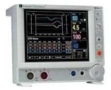
| Advanced monitors display complex data in simplified ways and interface with the bedside monitor for the display of additional parameters. |
Take away food, and, depending on the individual, a person can live for a few weeks or even months. Take away water, and a life is shortened to just a few days, a week at the most. Take away oxygen, and an individual will die within minutes.
Hypoxia, or low oxygen, will not result in immediate death, but it will have a significantly negative health impact that can affect a patient’s recovery. Subsequently, critically ill patients are monitored to ensure that oxygen delivery is maximized. An important component of this is cardiac output.
Oxygen is first absorbed through the lungs but delivered throughout the body in the bloodstream. “The cardiac output is a good parameter to tell how well the heart is doing in getting the blood out there,” says David Bailey, clinical engineer with the Mayo Clinic Arizona, Scottsdale/Phoenix.
There are a number of technologies, old and new, that are able to monitor cardiac output. Many facilities rely on one type with today’s gold standard being continuous cardiac output, which uses thermodilution and a pulmonary artery catheter. The concept behind the technology was developed more than a century ago, and the equipment tends to be simple and reliable. However, research has shown that the pulmonary artery catheter may be associated with an increased risk of infection as well as other potentially negative patient outcomes. As the issue is explored, newer technologies are emerging that focus on eliminating invasiveness and patient risk, but none have replaced the gold standard.
Even as physicians use fewer pulmonary artery catheters, the number of patients who need cardiac output monitoring is increasing. The figures are rising not because the application of continuous cardiac output has expanded, but rather because the patient population has grown. As the population ages, patients tend to be sicker and therefore in greater need of critical care. As conditions such as heart disease, obesity, and diabetes increase, so does the need for vital care. And as medical technology advances, more patients may also require monitoring. “We’ve started a heart transplant program, which has caused the need for more continuous cardiac output monitoring,” Bailey says.
As device inventories expand, biomeds may find themselves responding to more calls related to this equipment, but currently, inventories and the equipment’s reliability have kept the biomed workload in this area low. Bailey shares that the Mayo Clinic Arizona holds about eight thermodilution-based continuous cardiac output systems in its inventory. The Baylor Regional Medical Center, Dallas, and The Heart Hospital Baylor Plano have about 20, according to the facilities’ senior biomedical equipment technician, Carol Wyatt.
Despite the need for light maintenance, both sources suggest that the best way to maintain the equipment is to understand how it operates. “As with any unit, if you know how it works when it’s working correctly, then it’s easier to figure out what is wrong when it’s not working correctly,” Wyatt says.
Understanding the Why
Continuous cardiac output is based on principles developed by Adolph Fick in 1870. The theory suggests that the blood flow through an organ impacts the absorption or release of a substance, such as oxygen, and can be measured by comparing the substance values in the arterial and venous flows. The Stewart-Hamilton equation, developed in the 1890s, calculates cardiac output using a nontoxic dye injected into the venous system and withdrawn and measured on the arterial side. In the 1970s, doctors HJ Swan and William Ganz used thermodilution along with a special temperature-sensing pulmonary artery catheter to measure cardiac output.
This thermodilution method has become the clinical gold standard. “Thermodilution basically means there is an indicator or substance that is introduced into the blood in the right atrium and ventricle that changes the temperature of the blood,” says Barbara “Bobbi” Leeper, MN, RN, CCRN, clinical nurse specialist in cardiovascular services at Baylor University Medical Center in Dallas. “The amount of change in temperature over a short interval is measured, and a washout curve is created.” A modified Stewart-Hamilton equation uses the data to calculate cardiac output.
Thermodilution-based continuous cardiac output uses a modified Swan-Ganz catheter incorporating a thermal filament, proximal and distal lumens, and a standard thermistor. The thermal filament is placed between the right atrium and the right ventricle; the thermistor lies in the pulmonary artery. “This method has been pretty much the same since first introduced. The FDA approved the technology in 1992,” Leeper says.
Continuous cardiac output monitoring provides an alternative to intermittent or bolus methodology, which requires a health care professional to stand at the bedside and make the injections manually. The conversion from intermittent to continuous offers a number of advantages that include the elimination of inaccurate injectate temperature, inaccurate measurement of the injectate temperature, inaccurate injectate volumes, and the use of inaccurate computation constants; the influence of respiratory cycle averages into the value; and the reduction of clinician time required to perform the injections and calculations.1
“Clinicians are happy to get continuous output without having someone needed to do the bolus all the time,” Bailey says. “On occasion, I have seen them question the unit and go back and do an injectate and get a reading that way to give them a better understanding of why they are seeing the values they are questioning.” Sometimes, this reveals that the catheter must be changed due to clots or plaque buildup; other times, it reveals issues such as changes in the patient’s condition.
New Options
There are concerns, however, about the use of the pulmonary artery catheter. “This is really the result of a study published in the Journal of the American Medical Association in September 19962 that showed that patients with certain diagnoses who have pulmonary artery catheters in place had higher mortality rates than patients with the same diagnosis who did not have them in place,” Leeper says. The article prompted a consensus conference and literature review that concluded more research was needed, according to Leeper. Some of these studies are still under way, but until they are complete, continuous cardiac output using thermodilution and the pulmonary artery catheter remain the gold standard, particularly for patients who have the catheter inserted anyway.
Other options do exist but have not yet proven ideal. “Impedance methods have some value because they do not require any invasive connections to the body,” Bailey says. “But because they use electrodes, you can have artifact and changes in parameters that can be affected by motion or connectivity.”
Arterial pressure-based cardiac output technology provides another option, but is still invasive, although the arterial catheter is considered less so than the pulmonary artery catheter. Studies have produced different results comparing the two, with some researchers finding the arterial pressure-based cardiac output to be less accurate than the gold standard3,4 and others finding the newer technology to be promising.5
“Most facilities do use continuous cardiac output [using thermodilution and the pulmonary artery catheter], but the arterial pressure-based cardiac output is really emerging more and more,” Leeper says. “This has coincided with a decline in the number of pulmonary artery catheters that are inserted.”
An Eye on Maintenance
There is not, however, much difference in the equipment used for continuous cardiac output and arterial pressure-based cardiac output. “One requires an arterial line and the other a pulmonary artery catheter,” Leeper summarizes. The remaining components include the computer and optical module, the injectate set, and a cable that attaches to the catheter. The temperature gauge is present on the catheter. In many instances, as in the case of the Vigilance II by Edwards Lifesciences, Irvine, Calif, the catheter is specific to the technology.
Many systems do a self-test when they are powered on and hooked up for first use on a patient. “If the user gets through that self-test with no error messages other than the expected ones—such as ‘check catheter’ or ‘something is disconnected,’ which is expected because they don’t have it all hooked up just yet—then they’ll set the time and verify the monitor is OK by doing an in vitro calibration on the unit,” Bailey says. “That basically checks the optical module to make sure it’s in good working order.”
The equipment tends to be reliable, particularly newer models. “These are actually pretty good units,” Wyatt says. Preventive maintenance schedules on the equipment are determined by past performance. “We do a risk assessment annually and look at the equipment’s previous history,” Wyatt says. “If there have been a lot of problems, we’ll do a safety check annually. If we don’t see problems in the historical data, then our time is better spent on equipment with higher risk.”
To complete a safety check on a continuous cardiac output monitor, Wyatt will inspect the unit for physical damage, run it through a power-on self-test, and check the patient cable, optical module, and catheter. The use of known good cables and test catheters assists with this process. “You want to make sure you acquire a catheter from the department because that enables you to build a test kit and do the performance check,” Bailey says. “You must either convince them to give you one or include it as part of the purchase from the manufacturer.”
Wyatt has found that when problems arise, they are often due to a bad cable. “If there is no physical damage—the unit hasn’t been dropped or bumped—it could be a software issue but is more than likely a cable,” she says. Cable issues are handled in-house and involve simply switching the bad cable for a good one. “I try to have extra cables on hand so once we identify a bad cable it can be replaced immediately, and there is no downtime or very little,” Wyatt says.
Bailey has found that past problems have often been related to the optical module, a challenge that has been markedly reduced with a switch to the Vigilance II. “The optical module is a key part of it and a part that we have had problems with in the past,” Bailey says. “If a piece is going to fail, that seems to be where the failure can occur more often.” He adds that this problem could be related to the age of the equipment. “The newer models have been nearly trouble free,” he says.
Often, the manufacturer handles problems with the optical module or computer. “There aren’t a whole lot of serviceable parts on the optical module or even the monitor itself short of a knob going bad. Typically, we send it back,” Bailey says.
Or, the manufacturer comes to the site. “It’s dependent on what kind of service contract we have with the company of the continuous cardiac output and the monitor,” Wyatt says. “Some require you to send the unit in, and some come out to the site. If you’ve done the repairs many times it’s easy—reloading the software or replacing the main board.” However, the biomed wants to be sure this effort is not fruitless. “You don’t want to send something back that isn’t really having a problem,” Bailey says. By duplicating the issue or determining where the problem is, the biomed can guarantee that there is indeed a maintenance issue.
Troubleshooting is often learned on the job and through vendors who tend to share information about the products. “Manufacturers are great resources and often very helpful answering questions over the phone,” Wyatt says.
Users tend to be helpful as well, with biomeds reporting few user issues. “There have been a few times where I have gone up and shown a user that they have a cable in the wrong port or might not have run the test properly so we review it, but is it a big problem? No. The nurses are really good about using and taking care of them,” Wyatt says.
That might be because they recognize the importance of the measurement. Without the proper levels of oxygen, patient outcomes will not be positive: oxygen is life, and continuous cardiac output can ensure that life continues pumping through critically ill patients who need it most.
Digging for Gold
Today’s preferred method for monitoring continuous cardiac output uses thermodilution and a pulmonary artery catheter. However, the medical community is no longer certain that use of the pulmonary artery catheter is the best way to treat patients. Concerns include an increased risk of infection, artery ruptures, air embolisms, and associated negative outcomes, including fatality. This has led to the exploration of alternate methods, though none have yet been declared the new “gold standard.”
Barbara “Bobbi” Leeper, MN, RN, CCRN, clinical nurse specialist of cardiovascular services at Baylor University Medical Center, Dallas, finds that arterial pressure-based cardiac output technology is emerging “more and more.” The typical method, however, is still considered invasive; the technology requires access to the radial or femoral artery using a standard arterial catheter. The FloTrac system by Edwards Lifesciences, which uses this method, calculates cardiac output using stroke volume and pulse rate generated by the left heart compared to the direct cardiac output measurements of the right heart taken by the thermodilution technology. Noninvasive pulse pressure methods are based on similar principles but use a sphygmomanometer or tonometer to measure pulse pressure through the skin. Unfortunately, these methods do not tend to be as reliable.
Doppler methods generally provide a noninvasive technique for monitoring blood flow and cardiac output. Most Doppler methods measure blood flow and volume through detection of the resulting Doppler shift; these measurements are then used to calculate cardiac output. Doppler ultrasound uses ultrasound to read the measurements directly in the heart; echocardiography uses echocardiographic measurement of blood flow to obtain the measurements; and ultrasonic cardiac output monitoring uses an external ultrasonic transducer element that employs continuous wave Doppler to determine the flow profile. Esophogeal Doppler or transoesophogeal Doppler is an invasive Doppler method that uses a probe inserted into the patient’s esophagus and placed at the midthoracic level to measure blood velocity in the descending aorta for use in cardiac output calculations.
Impedance methods are completely noninvasive and determine cardiac output through volumetric changes measured by the corresponding changes in thoracic impedance. This is often referred to as impedance cardiography or thoracic electrical bioimpedance. David Bailey, clinical engineer with the Mayo Clinic Arizona in Scottsdale/Phoenix, notes the systems are valuable because they are noninvasive, but the use of electrodes can lead to artifacts resulting from motion or connectivity.
Because of their imperfections none of these methods has yet replaced today’s preferred method, but that does not prevent researchers and clinicians from digging for gold.
—RD
Renee Diiulio is a contributing writer for 24×7. For more information, contact .
References
- Edwards Lifesciences LLC. Continuous cardiac output. www.edwards.com/presentationvideos/powerpoint/cco/cco_speaker_notes.pdf. Accessed May 1, 2008.
- Dalen JE, Bone RC. Is it time to pull the pulmonary artery catheter? JAMA. 1996;276(11):916-918.
- McGee WT, Horswell JL, Calderon J, et al. Validation of a continuous, arterial pressure-based cardiac output measurement: a multicenter, prospective clinical trial. Crit Care. 2007;11(5):R105.
- Prasser C, Bele S, Keyl C, et al. Evaluation of a new arterial pressure-based cardiac output device requiring no external calibration. BMC Anesthesiol. 2007;7(1):9.
- Lemson J, van der Hoeven JG. Clinical value of an arterial pressure-based cardiac output measurement device. Crit Care. 2008;12(1):403.


