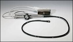
The Seno Imagio breast imaging system and the associated predictive model may significantly improve a physician’s ability to accurately rule out breast cancer compared to traditional ultrasound alone, according to manufacturer Seno Medical Instruments.
“Diagnostic specificity, or the ability to accurately identify benign masses, remains disappointingly low for imaging methodologies optimized to identify all cancerous lesions with near 100% sensitivity,” said A. Thomas Stavros, MD, medical director, Seno Medical Instruments. “We believe that by training Imagio readers with this real-time predictive model, they may be able to accurately reclassify benign breast lesions to a lower BI-RADS score so the patient can confidently avoid a biopsy on benign masses. If confirmed by Seno’s prospective, multicenter PIONEER Pivotal Study of Imagio, the predictive model may improve the image reader’s ability to accurately characterize solid breast masses as cancerous or benign and to spare women with benign lesions from the biopsy process beyond the standard-of-care today.”
The predictive model is based on key optoacoustic features of breast masses obtained by Imagio during a 79 subject pilot study. Seno completed active enrollment of 2,100 subjects in the U.S.-based PIONEER study in September. The results of the study will serve as the basis for the company’s Premarket Approval Application (PMA) with the FDA.
To develop the real-time predictive model, a radiologist blinded to histologic outcomes evaluated traditional diagnostic breast ultrasound and the 5 different Imagio optoacoustic features of 79 masses (41 benign, 38 cancer) classified BI-RADS 4 prior to biopsy. Linear regression was used to model and predict the probability of malignancy, while logistic regression was used to model and to predict whether a mass was benign or malignant.
Imagio was designed to identify two functional hallmarks of a potential malignancy: the presence of abnormal blood vessels and the relative reduction in oxygen content of hemoglobin. The technology is non-invasive and does not require contrast agents or radioisotopes, nor does it use ionizing radiation (x-ray).


