Royal Philips named new milestones in the development of its spectral detector angio CT solution — Philips Spectral Angio CT suite — which aims to bring the company’s spectral CT imaging technology into an integrated hybrid angio CT suite.
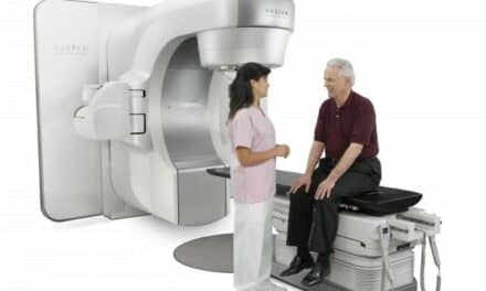
By combining its Spectral CT 7500 system [1] and its Image Guided Therapy System – Azurion with FlexArm – in a fully integrated interventional suite solution, Philips aims to give physicians immediate access to these two key imaging modalities in a single room, enabling innovation in minimally-invasive procedures in areas such as oncology, stroke, and trauma care.
Philips announced a new clinical partner and highlighted clinical studies that focus on the added value of using spectral CT imaging technology during interventional procedures.
Expanding Philips’ Clinical Network
Leiden University Medical Center (Leiden, the Netherlands) has joined Philips’ global network of clinical partners to investigate how its spectral detector angio CT solution could potentially offer new treatment opportunities and improve patient care.
“We are excited to co-create an innovation that could play a defining role in improving patient care in the space of interventional oncology,” says Mark Burgmans, MD, head of Interventional Radiology at Leiden University Medical Center. “Adding spectral CT imaging to the interventional suite will enable us to offer new treatment opportunities, avoid moving patients from one imaging suite to another, and offer the unique benefits of spectral CT information when you need it.”
Other leading clinical institutes that Philips is working with on this innovation are Mayo Clinic and Baptist Health’s Miami Cardiac & Vascular Institute.
Philips Spectral Angio CT suite combines the company’s latest diagnosis and treatment technologies. Philips Image Guided Therapy System – Azurion with FlexArm – is the company’s next-generation image-guided therapy platform, integrating imaging systems, software, and specialized diagnostic and therapeutic devices to support exceptional treatment for the most complex procedures. The addition of Philips’ award-winning Spectral CT 7500 system means physicians only need one scan to capture all the spectral information required to differentiate and quantify different tissues.
Spectral CT enables improved detection, delineation, and quantification of lesions, leading to better-informed planning for minimally-invasive procedures and more precise interventions. This technology has demonstrated higher sensitivity in detecting malignant findings and improved readings of incidental findings [2][3].
Through continuous research, Philips is building clinical evidence that supports the added value of spectral CT imaging for diagnosis and treatment guidance.
Research Results
At this year’s Cardiovascular and Interventional Radiological Society of Europe Annual Meeting is a presentation being given by Filippo Piacentino, interventional radiologist at the University of Insubria, on the value of spectral CT imaging guidance for performing high-confidence tumor biopsies [4]. The results being presented illustrate the potential for Philips’ spectral CT technology to better guide biopsies by distinguishing between active and non-active regions in a tumor. Ensuring that a biopsy contains a high number of actively dividing cancer cells is important for high-confidence diagnosis.
“With conventional CT, large masses may appear as a largely uniform mass, making highly targeted biopsy difficult,” says Piacentino. “By fusing images from Philips’ XperGuide live needle guidance with images from spectral CT, that are color-coded based on the effective atomic number of tissues and provide a large amount of additional information, we can now investigate the possibility of obtaining better defined biopsy targets with a fewer number of inconclusive biopsies.”
References
[1] Minnie Award for Best New Radiology Device
[2] Analysis by Aarhus University Hospital Aarhus, Denmark. Results from case studies are not predictive of results in other cases. Results in other cases may vary.
[3] Analysis by University Hospital Cleveland, USA. Results from case studies are not predictive of results in other cases. Results in other cases may vary.
[4] Filippo Piacentino, ‘Spectral CT as innovative imaging guidance in large lesions lung biopsies. XperGuide and Z-effective fusion for more defined targets, more diagnostic samplings and more biomarkers information’. CIRSE 202