Alternating Pressure Support Surface Launched for Perioperative Pressure Injury Prevention
The low-profile overlay is designed for use during extended surgeries to reduce the risk of hospital-acquired pressure injuries.
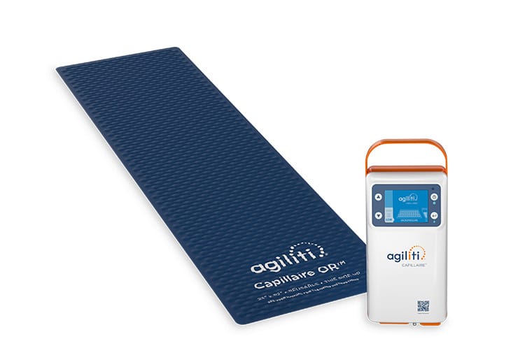
The low-profile overlay is designed for use during extended surgeries to reduce the risk of hospital-acquired pressure injuries.
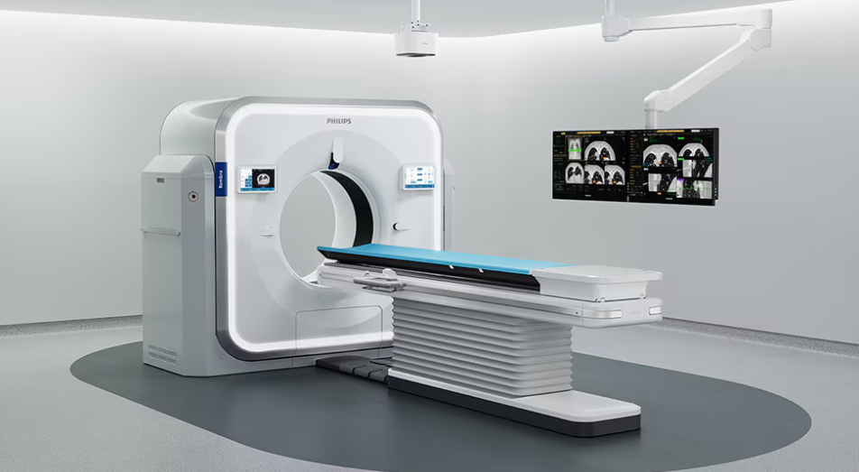
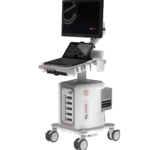
The new CT platform features fast reconstruction speeds, an 85-cm bore, and AI-enabled workflows aimed at improving throughput in acute care environments.


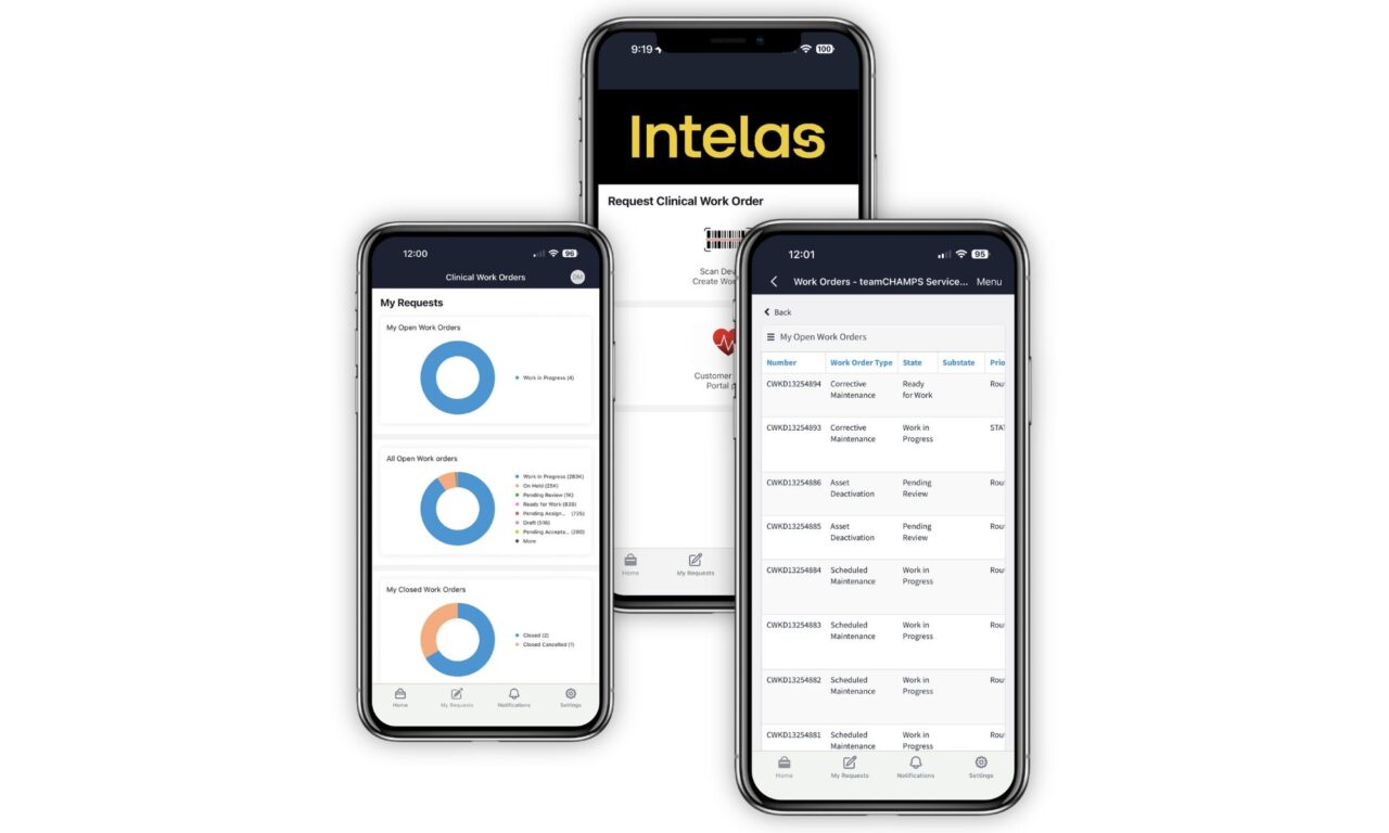
The new mobile experience connects technicians, clinical staff, and hospital leaders on a single platform to help accelerate issue resolution and improve visibility across medical equipment service.


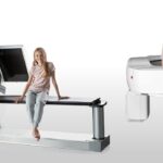
The low-profile overlay is designed for use during extended surgeries to reduce the risk of hospital-acquired pressure injuries.

Routine electrical safety testing is frequently defended on instinct rather than evidence. An engineering analysis shows what actually protects patients and staff.

The $35 million expansion will advance artificial intelligence-powered ultrasound solutions for trauma assessment and mass casualty incident preparedness.

The expanded partnership brings integrated patient monitoring technologies into outpatient settings as more procedures shift away from hospitals.
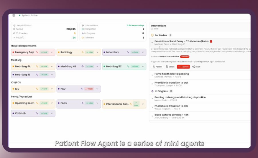
The platform combines real-time location systems and EHR data to optimize patient flow, equipment management, and clinic scheduling without requiring new infrastructure.
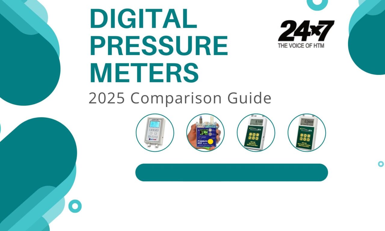
Compare digital pressure meters used to measure gas, liquid, and air pressure in medical equipment, with key specs and features highlighted.
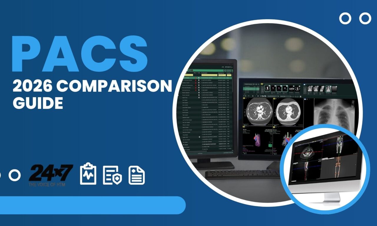
A side-by-side look at PACS capabilities, from supported modalities and interoperability standards to pricing, licensing, and EHR integration.
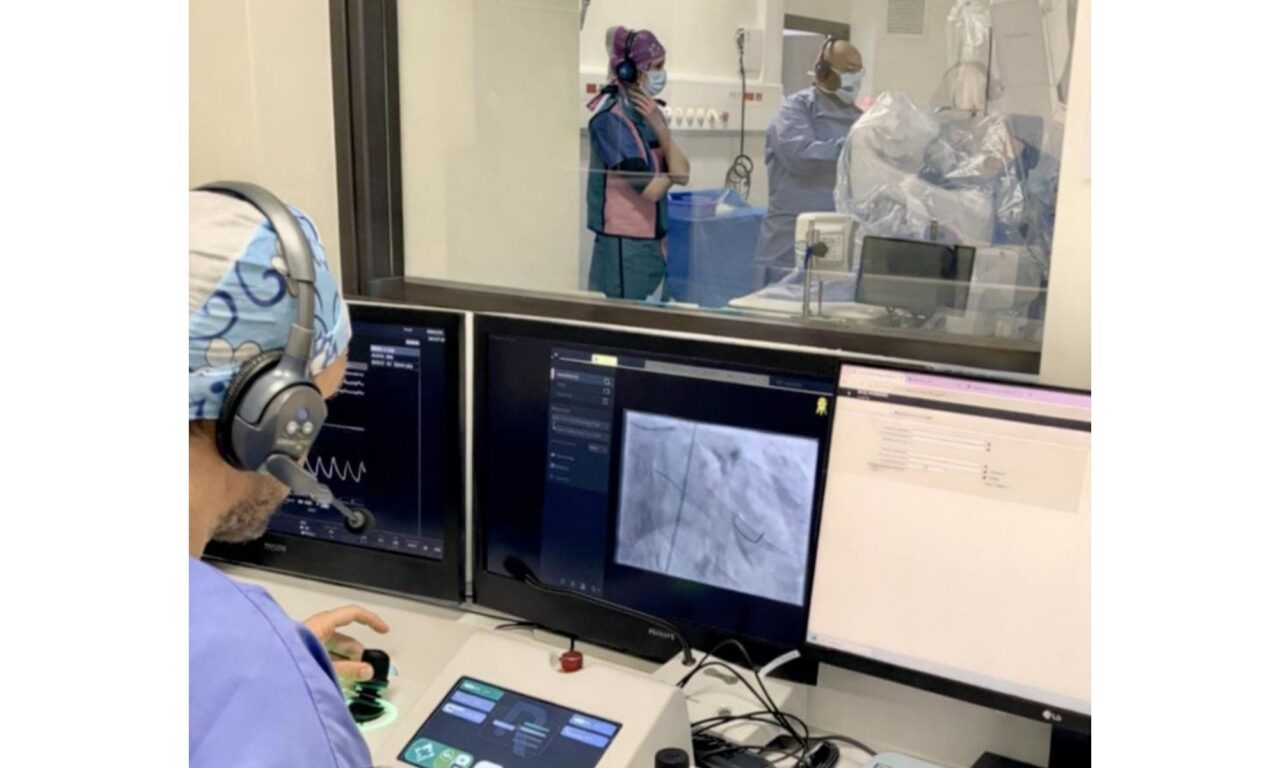
The study will evaluate a second-generation robotic platform in complex catheterization lab procedures.
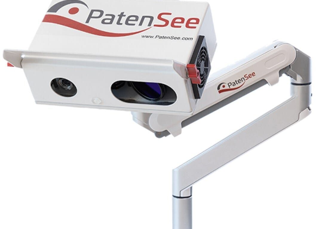
The AI-driven optical system is designed for early detection of stenosis in hemodialysis patients and to reduce clinical staff workload.

The acquisition aims to integrate Armis’ real-time asset discovery and cyber-physical security capabilities into ServiceNow’s security workflows.
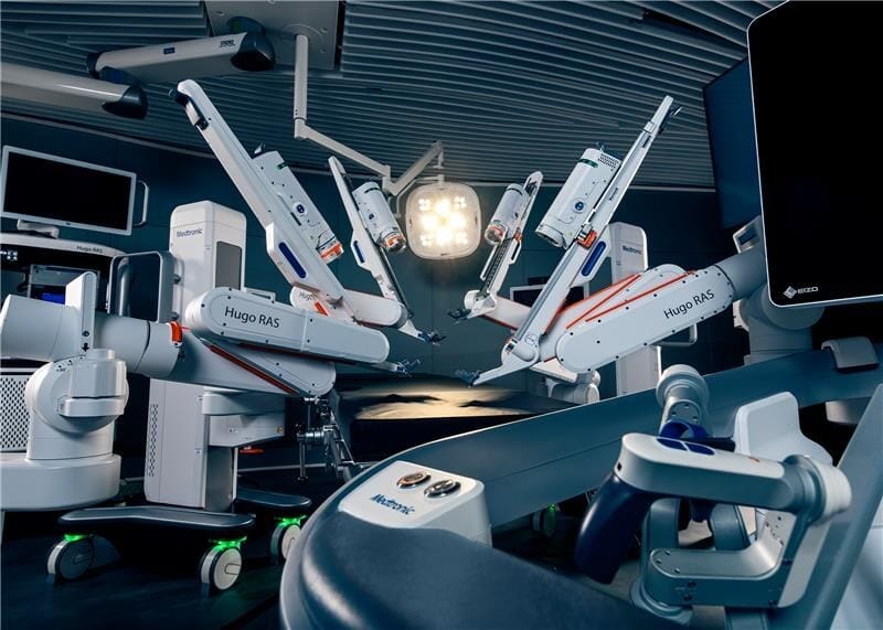
The system is indicated for use in minimally invasive urologic surgical procedures including prostatectomy, nephrectomy, and cystectomy.