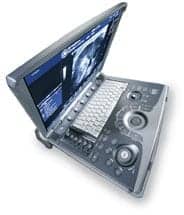
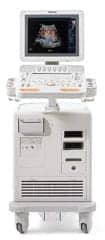
In the 21st century, your first baby picture is likely to be snapped in the womb, and the same with the first video. Ultrasound exams—two-dimensional, three-dimensional, and four-dimensional—have become routine steps in prenatal care. With today’s image quality, parents who do not want to know the sex of their baby may find it difficult to ignore. But ultrasound does a lot more than capture a child’s earliest days.
As the technology has advanced, ultrasound has become a significant diagnostic tool. Improved image quality and greater accuracy have led to more applications for the modality and greater volumes of associated testing. The increased use has driven the expansion of ultrasound inventories as well as the evolution of service solutions.
Many health care institutions have brought as much ultrasound service in-house as possible. The economies of scale justify the move, and the savings—particularly in equipment uptime—provide a huge benefit. However, a biomedical/clinical engineering department can find it challenging to obtain the software, training, parts, and/or time needed to optimize device performance and uptime solely on its own.
Flexibility, innovation, and education are often part of the solution. And where a biomed program leaves off—very often with the probes—a manufacturer or third-party service picks up. Walter Barrionuevo, director of clinical engineering services at BayCare Health System, based in Clearwater, Fla, describes a typical arrangement: “The majority of ultrasound units are serviced by our in-house technicians with backup assistance from the OEM,” Barrionuevo says. “A service arrangement with a transducer repair company provides us with loaners whenever one of our transducers requires service.”
In addition to loaners, trusted service providers may also provide repairs, maintenance, and parts at a less expensive cost then the OEM. They may also be able to respond more rapidly. “Everyone needs a company they trust that they can call for a loaner and have it there the next day,” says Elaine L. Freeman, CBET, biomedical support services for FirstHealth of the Carolinas in Pinehurst, NC.
Loaned probes ensure that an ultrasound department can continue to see patients while equipment is out for repair. Because of the sensitive nature of probes, they are very often sent out rather than serviced in-house. However, in-house maintenance, first look, and care policies can help to extend the life and uptime of a probe.
 |
| As the technology has advanced, ultrasound has become a significant diagnostic tool. Improved image quality and greater accuracy have led to more applications. |
Tackling Time
Preventive maintenance (PM) can help to maximize ultrasound equipment, but often, it will take the complete breakdown of a device to make it available to the biomed.
“Technologists don’t want to give the machine up with a minor problem. Unless the system doesn’t come up all the way, they won’t give it to us,” says Chris Jones, Sr, MCP, CPACS associate, lead imaging service technician in the imaging services division of the clinical engineering department at Johns Hopkins Bayview Medical Center in Baltimore.
This means biomeds need to be flexible in performing PM. “I have to flex my hours in order to do my 6-month preventive maintenance on those machines,” Freeman says, who oversees an ultrasound inventory of about 25 units. She notes that as a result of their busy use, much ultrasound maintenance is handled on a “crash-and-burn” basis.
Even so, users still benefit from greater uptime. “All the user has to do is call, and hopefully, I can come and fix the machine as quickly as possible. They don’t have to wait 12 or 24 hours to get back up,” Freeman says.
To save time, Freeman keeps a physical notebook on every device in which she stores all relevant data, including manufacturer and contract information, IP addresses, maintenance history, parts information, and preset disks. Whenever there is a problem, she brings the device’s notebook along. Computerized equipment maintenance systems can also keep this information, with digital benefits such as retrieval from anywhere within a facility. However your system works, though, the point is quick and easy access to the data, which can save a biomed both time and frustration.
“Biomeds want to know their machines, the revisions they are running, their IP addresses, and anything else that can help to solve a problem,” Freeman says. “The main thing is to have all the information needed to put the machine back together.”
Getting Trained
Much of this information is learned during training and is considered imperative to delivering service. “I would suggest [to a biomed department] bringing ultrasound service in-house that at least one person be manufacturer trained,” Freeman says. Freeman has been to GE and Siemens training schools to learn how to service the devices FirstHealth has purchased over the years.
Today, manufacturer training is typically negotiated with the original purchase agreement, but it can also be obtained later—although often at greater expense. Third-party educators can provide a less costly and sometimes easier option.
“The manufacturer wanted me to pass a test on basic ultrasound before enrolling in the training course. A third party won’t make you jump through all of those hoops,” Freeman says.
Of course, some knowledge of ultrasound and scanning is necessary to be able to troubleshoot. “Biomeds should have an understanding of what ultrasound does, how it works, and what to do with it,” Jones says.
Coursework often includes self-scanning as a machine diagnostic tool. “They teach you to scan your jugular or your liver—the easiest [anatomy],” Freeman says.
Although it may be interesting, Freeman questions exactly how useful this self-scanning is since many problems are tied to the software. “The image is not going to degrade, like in the old days,” Freeman says. “It either works or it doesn’t.”
Software Solutions
Software is a common problem, but the frequency tends to vary according to equipment make and model, says David Bishop, MCP, a BMET III in the clinical engineering department of Mease Dunedin Hospital, Dunedin, Fla. “Corrupted software is a prominent issue in some systems. In one, problems seem to be generated by corruption due to improper shutdowns. In another, the system software may not run normally due to simply being bogged down with excess patient files. I’ve seen other software issues as well,” Bishop says.
Generally, software issues are easily solved—”assuming you have proper service access to the equipment,” Bishop adds. “In some cases, just getting access to viable training or service passwords can be an issue.”
In an effort to protect proprietary data, manufacturers can sometimes be stingy with information—some worse than others. Freeman notes that she has gotten the software for every machine in her inventory, but other biomeds with equipment from other manufacturers report little to no success in this area.
The situation can be frustrating. The lack of information can negatively impact a biomed’s ability to properly diagnose and fix a problem as well as increase the cost of service. “We have to pay exorbitant labor prices to the manufacturer to reload the software,” Barrionuevo says.
On the contrary, with access to the software, biomeds can typically solve related problems themselves and avoid this cost. Jones recommends biomeds know the software, its level, and have a backup. “If you have a network configuration, have that loaded too,” Jones says.
Software reloads are often an easy and quick answer. “If we suspect a software problem, we like to reload because it’s easier to go back to ground zero than to try to troubleshoot and fix the software piece that is problematic,” Jones says. “If we go back to zero with the factory install, it solves 95% of the problems.”
 |
| The lack of service information can negatively impact a biomed’s ability to properly diagnose and fix a problem. |
Hardware Hardships
When software is not the problem, hardware is; in that case, the challenge becomes an economic one. “A bank of spare parts is out of the question when just about every board is $6,000 to $10,000,” Freeman says. If by chance the wrong part is ordered, restocking fees can be 30% to 35% of a part’s cost.
Biomeds can realize significant savings by purchasing parts from authorized service and repair centers, particularly after warranties have expired. “When a unit reaches end-of-service life and is no longer supported for parts by the OEM, many of these repair/service centers will extend most parts support for years,” Bishop says.
Without spare parts, troubleshooting could take longer. Freeman gets around this by swapping parts with an identical machine that is working. “The beauty of having more than one of the same machine is that you can roll them side by side and swap parts to figure out what is really giving you the problem,” Freeman says.
Although the technology has advanced, the units’ parts are still basically replaceable to a module/card level, Bishop notes. Probes are also an easy switch, but equally expensive. “You can pay in excess of $3,000 for the repair of a standard linear or curved probe through an authorized repair center,” Bishop says. “This amount can easily double if repaired at an OEM’s repair facility,” adding that transesophageal echo (TEE) and transvaginal probes are significantly more expensive to repair or replace than standard linear or curved probes.
Probe Problems
Unfortunately, it is the TEE and transvaginal (or intravaginal) probes that are the most problematic. “Physical damage is the most frequent issue. Otherwise, it would be component failure due to moving internal parts,” Bishop says.
Bishop describes the transvaginal probes as longer and more cumbersome than standard linear or curved probes. Too much pressure can introduce cracks during an exam, a whipcord accident, or a fall to the floor.
TEE probes are similarly vulnerable. “TEE probes are susceptible to patients involuntary biting as the probe is introduced into the esophagus, damaging the probe housing,” Bishop says. Bite guards help to avoid this damage.
Both probes, which are used internally, require powerful sterilization that often takes the form of a Cidex soak. “That can introduce fluid into the probe if any pinhole is present in the membrane and do damage to the internal components,” Bishop says.
Probes should therefore undergo a thorough exam before sterilization, a procedure that happens with differing regularity depending on the institution, department, and personnel.
“Users can present service challenges by their treatment of the equipment and probes. Training the department staff on proper treatment of the equipment has helped,” Bishop says. However, training can have different results.
An ultrasound group at FirstHealth who handles the intravaginal probes still tends to oversoak the devices, despite training, resulting in three or four failures per year. However, central processing at the same facility, which took over the sterilization of TEE probes, has contributed to a decrease in damage to those instruments through procedural compliance, according to Freeman.
Jones has had a similar experience. In one section of Johns Hopkins Bayview Medical Center, probe repairs have run roughly $70,000. In another, notably the departments of radiology, vascular, and echocardiography, the technologists are much more careful and the costs have been much less. “I think in the past 5 years, among those three entities, the most I’ve spent is $20,000. In radiology, I’ve spent only $5,000 in 5 years,” Jones says.
Care Instructions
User training elements are simple: “Be careful not to drop the probes. Only soak transvaginal or TEE probes as long as is called for. Hang slack cable on the unit’s designated hangers,” Bishop says. Additional tips include consistent use of bite guards with TEE probes and PM on all equipment.
“How often a PM is accomplished would be per the manufacturer and the facility’s recommendation/policy. My preventive maintenance procedure would include a physical inspection, electrical safety inspection and leakage test, and inspection of certain image details using an approved phantom,” Bishop says.
PM is performed in-house, but repairs are sent out. Probes are complicated devices containing delicate crystals, and problems are best fixed at a repair center. “They have to be sent out to a qualified repair center. As they are complex, few repair centers can repair these probes,” Bishop says.
Third-party firms are often much less expensive than the manufacturer. Most biomed departments shop around to find a company that can offer savings but maintain quality. “We’ve shopped around to find out where we can do repairs for less cost than an exchange with the OEM,” Jones says. To ensure quality, Jones has visited his third party’s facilities, where probes are stripped and rebuilt.
Partners can be just as important as parts. Freeman recommends maintaining friendly relations with the OEM as well as third-party providers. “Get to know your field service person and become their friend. Sometimes that goes a long way,” Freeman says.
With ultrasound increasing its value as a diagnostic tool, more biomeds may want friends in provider places. Many find themselves in similar positions, struggling with time, training, budgets, and policy issues so that they can maximize the uptime and performance of ultrasound devices. But their flexibility and innovation ensure that doctors get the diagnostic data they need and parents still get that first snapshot of baby in the womb.
Renee Diiulio is a contributing writer for 24×7. For more information, contact .
Ultrasound Performance and Utility
Ultrasound is like sonar—they both operate on the same principles. The probe (or transducer) is the submarine or bat, and the anatomy is the enemy or insect.
As the sonographer places and moves the transducer, it emits sounds waves that are reflected by anatomical structures. As they bounce back, they are analyzed by the computer software to capture still and moving images of inner anatomy—without having to open the body.
Operating frequencies for ultrasound fall in the 1 MHz to 50 MHz range, with 30 MHz to 50 MHz representing high-frequency systems. (The upper range of human hearing tops out at 20 KHz). Lower frequencies are more often used by external systems.
Older ultrasound systems produced 2D sectional images; newer systems offer 3D and 4D (motion) imaging options. Doppler ultrasound instruments, first developed in the 1960s, visualize blood flow, targeting red blood cells with the sound waves. The common techniques tend to differ with the department. For instance, Doppler and 4D technologies are used most commonly in cardiac and obstetric/gynecological units.
As ultrasound technologies have advanced, greater power, improved image quality, and smaller components have driven an expansion in the already broad application of the image modality. An ultrasound exam may be ordered to gather information about many anatomical structures, including the bladder, eyes, liver, gallbladder, heart and blood vessels, kidneys, ovaries, pancreas, spleen, thyroid and parathyroid glands, scrotum (testicles), uterus, and the fetus in pregnant patients.
Exams often require a specialized probe, designed to effectively elicit the necessary image needed for a diagnostic. For instance, the transesophageal—or TEE—probe is used to examine the heart and valves from an internal position reached through the throat. A transvaginal (or intravaginal) ultrasound examines female reproductive organs; the transvaginal probe provides a “closer” look from inside the woman’s vagina.
The shape of the probe creates the field of view, and the frequency used impacts penetration and resolution. Sonographers are trained in the software and probe’s use and are responsible for the acquisition of the images.
Recent developments include advances in image quality and resolution, 3D and 4D applications, and greater portability. Future developments are likely to include more of the same, resulting in even further use and growth.
—RD


