The low-dose X-ray technique shows how shoulder mobility differs after two surgeries, giving doctors new data to guide treatment decisions.
A new study by Emory Healthcare researchers uses dynamic digital radiography (DDR) to compare shoulder biomechanics in patients after two widely accepted surgical interventions for massive irreparable rotator cuff tears (MIRCTs).
Reverse shoulder arthroplasty (RSA) and arthroscopically assisted lower trapezius tendon (aLTT) transfer are often used to repair these tears; however, quantifying shoulder function post-operatively has remained difficult to assess. However, with the use of novel DDR imaging, it provided the ability to examine in-vivo kinematics by measuring scapulohumeral rhythm (SHR), the ratio of the glenohumeral and scapulothoracic contributions to shoulder motion, non-invasively in patients.
The authors also aimed to design an objective methodology for selecting the appropriate intervention that will maximize the patient’s shoulder mobility with the help of DDR. The study is published in the Journal of Shoulder and Elbow Surgery.
DDR: A Low-Dose X-Ray Imaging Technique
DDR is a novel, low-dose X-ray imaging technique available on Konica Minolta Healthcare DR Systems that captures both static images and cinegrams, providing an innovative way to obtain detailed images of complex joints like shoulders while in motion. By acquiring a series of images at high speed, DDR generates a cineloop that enables clinicians to visualize anatomical motion over time (cineradiography), enhancing the system’s diagnostic capabilities.
Utilizing DDR to characterize scapulohumeral rhythm both pre- and post-operative and evaluate for precise changes in SHR, Sameer R. Khawaja, MD, and collaborators, including the leadership of Eric R. Wagner, MD, MSc, and his research lab, demonstrate that Wagner’s patients undergoing aLTT yields superior restoration of shoulder biomechanics for patients with massive irreparable rotator cuff tears than his patients undergoing RSAs.
Rotator Cuff Surgery Outcomes Compared
The study highlights how aLTT transfer enhances shoulder stability and improves functional mobility. In contrast, RSA is a very successful treatment of massive irreparable rotator cuff tears and other pathologies but fails to restore the same level of native biomechanics as the aLTT.
“Using the dynamic radiography provided by Konica Minolta’s DDR imaging enables us to change the clinical algorithms for both preoperative decision making and postoperative evaluations of surgical outcomes,” says Wagner in a release.
Zaamin Hussain, MD, an orthopedic surgery resident at Emory Healthcare, adds in a release, “It’s often difficult to decide whether we should do a RSA or aLTT for these patients with massive rotator cuff tears. These are very different treatments. Not only can the DDR images help make that decision preoperatively, but the results of this study suggest there is potential for improved overall coordination of the shoulder with aLTT, which is only possible to assess with in vivo dynamic imaging.”
DDR Enhances Rotator Cuff Surgery Outcome Analysis
John Sabol, PhD, clinical research manager at Konica Minolta Healthcare, says, “Konica Minolta congratulates the team at Emory Healthcare on the publication of their study demonstrating the clinical utility of DDR in comparing post-surgical outcomes in patients undergoing reconstructions for massive irreparable rotator cuff tears. DDR is an FDA-cleared radiography solution that provides insight into the dynamic relationship of bones and soft tissue through their full range of motion.
“As the Emory team has demonstrated in this work, DDR overcomes the historical challenges in evaluating biomechanics in clinical patient populations. This will enable improvements in the quality of care and patient outcomes.”
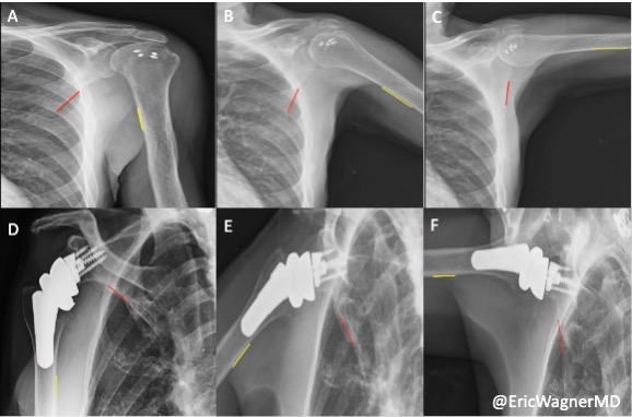
Photo caption: Radiographs of humerothoracic abduction in patient with aLTT (top) and RSA (bottom) at: (A and D) rest, (B and E) 45°, (C and F) and 90°. aLTT, arthroscopic-assisted lower trapezius tendon; RSA, reverse shoulder arthroplasty.
Photo credit: Konica Minolta Healthcare Americas

