Exclusive to the Sonosite LX system, the new transducer aims to improve diagnostic confidence and procedural precision in superficial imaging.
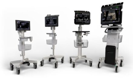
Fujifilm Sonosite Inc introduced the UHF46-20 transducer, a 46MHz ultra-high frequency transducer for point-of-care ultrasound.
With a minimum scan depth of 4mm, the UHF46-20 transducer is designed to enable clinicians to clearly visualize the first one to two centimeters beneath the skin and identify structures smaller than 1mm, such as superficial nerves and vessels, with high-quality resolution. The transducer is built upon the ultra-high frequency technology from Fujifilm VisualSonics and available exclusively on the Fujifilm Sonosite LX POCUS system.
“The UHF46-20 transducer, when paired with the Sonosite LX system, enables the largest frequency range of any point-of-care ultrasound system on the market today, addressing a longstanding challenge that current clinical ultrasound systems have been unable to overcome,” says Richard Fabian, president and chief executive officer of Fujifilm Sonosite Inc, in a release. “We’re proud to bring to market the UHF46-20 transducer as the first and only 46MHz UHF transducer in [point of care ultrasound] to provide clinicians with an unparalleled tool that may help them enhance diagnostic confidence and procedural accuracy.”
The combination of the new transducer and the Sonosite LX offers clinicians a solution for a spectrum of imaging needs, spanning from deep abdominal scans to superficial assessments. The UHF46-20 Transducer holds promise for improving outcomes across a range of sensitive applications including neonatal intensive care unit (NICU) and rheumatology.
In the NICU, its resolution may help clinicians see superficial submillimeter anatomy that conventional ultrasound may not capture. The use of ultra-high frequency ultrasound may aid clinicians in improving procedural quality by allowing for better visualization of tiny anatomy that may help to improve first attempt success rates.
In rheumatology, ultra-high frequency’s superficial imaging may aid in the visualization of subclinical synovitis, erosions, crystal deposits, and inflammation, all of which are beneficial in the early diagnosis and intervention of chronic conditions.
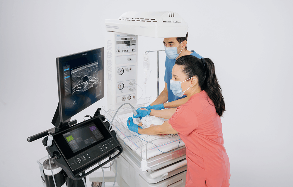
Photo caption: UHF46-20 transducer used with the Fujifilm Sonosite LX POCUS system
Photo credit: Fujifilm Sonosite