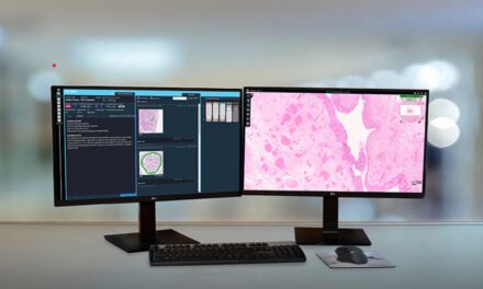
SpectraWAVE announces U.S. FDA 510(k) clearance for product enhancements to the HyperVue Imaging System. The intravascular imaging system combines DeepOCT images and near infrared spectroscopy (NIRS) to support physicians optimizing coronary stenting in the cardiac catheterization lab.
Newly cleared product enhancements include contrast-free saline imaging, artificial intelligence algorithms to support the identification of key clinical structures of calcium and external elastic lamina (EEL), and hands-free angiographic co-registration.
The latest clearance adds to the existing HyperVue toolkit, which includes artificial intelligence enabled lipid, lumen, stent, and sidebranch detection, as well as the no-flush prep Starlight Imaging Catheter designed for efficient setup and image acquisition in complex lesions.
“Intravascular imaging is essential to optimize patient outcomes,” says Richard A. Shlofmitz, MD, chairman of cardiology of the St. Francis Hospital and Heart Center. “I use intravascular imaging in all my coronary stenting patients, and I’m delighted to see SpectraWAVE enter the arena and push the limits of these technologies to help us better care for our patients.”
He adds, “The combination of DeepOCT and NIRS is ground-breaking and provides important clinical information that was previously unavailable. With the recent updates that allow for contrast-free saline imaging and support image review with artificial intelligence, this product will provide significant value to both experienced physicians and those just beginning to embrace imaging.”
Eman Namati, PhD, CEO of SpectraWAVE, also spoke out about the clearance, commenting: “Optimal stenting outcomes for patients, as demonstrated by the wealth of randomized clinical studies and meta-analyses, are predicated on the ability to acquire high-quality intravascular images, and distill the data into clinical actions. “We have pushed the limits of image quality with our DeepOCT-NIRS technology, created a simple low-profile no-flush prep catheter, and enabled long and fast pullbacks.”
What’s more, Namati says, “We have now expanded our artificial intelligence algorithms to support EEL-based vessel measurements, fully automated angiographic co-registration to map findings back to the angiogram, and enable saline imaging to mitigate the need for additional contrast use.”
Intravascular imaging is a tool to optimize coronary stenting procedures, providing key insights into plaque morphology, plaque modification decisions, stent and balloon sizing and landing zone selection, confirmation of treatment optimization, and future adverse event risk.