A review of some of the common devices used for eye examination and diagnosis and problems biomeds might encounter when maintaining and servicing them.
Ophthalmologists and other eye care professionals use many devices to diagnose and treat eye problems. Part I of this article will review some of the common devices used for examination and diagnosis. Part 2 will review devices used for the treatment of eye problems.
Most ophthalmic diagnostic devices have optical components, such as lenses, mirrors, and prisms. Many of these components have a special thin coating for filtering specific wavelengths of light, for reflecting light, or for reducing reflection. Great care must be exercised when removing dust and stains on optical components to avoid scratching or removing the surface coating. Dust and stains become harder to clean when they accumulate and therefore periodic cleaning is recommended. Excessive cleaning can lead to quick deterioration of the surface coating, so manufacturer instructions for frequency and method of cleaning of each device should be followed. All ophthalmic equipment should be kept under dust covers when not in use.
Bulbs are also common in many ophthalmic devices. When replacing bulbs, care should be taken to not touch them with bare fingers. Oils from the skin create hot spots on the bulb that can shorten the bulb’s life. Additionally, fingerprints can become etched into the bulb’s glass jacket and cause a shadow on the illumination field.
Direct Ophthalmoscope
A direct ophthalmoscope is a handheld instrument for routine examination of the inside of the eye. It contains a battery, a variable light source, and a set of lenses used to focus on particular structures of the eye. The device is held in front of the patient’s eye and the operator looks through one of the small lenses into the eye to view the appearance of the cornea, the lens, the aqueous and vitreous humor, and the surface of the retina. The view provided by the ophthalmoscope is monocular, nonstereoscopic (2-D), narrow field (5°), and its magnification is approximately 15x.
Indirect Ophthalmoscope
A binocular indirect ophthalmoscope (BIO) is worn as a headset and is used in conjunction with a condensing aspheric lens held against the patient’s eye. A BIO provides a much wider field of view (45°) than a direct ophthalmoscope and permits viewing of almost all of the patient’s retina. The BIO is the viewing instrument of choice for retinal examinations. The view provided by the BIO is stereoscopic (3-D), inverted, and illuminated with magnification of about 5x. Some BIOs have a built-in video camera to permit eye care professionals in training to view the examination on a screen.
Slit Lamp
A slit lamp is a device designed for examination of the external and internal anterior structures of the eye. Eye care professionals use slit lamps to identify diseases, spot foreign bodies, fit contact lenses, and visualize surgical laser procedures. The slit lamp is composed of a microscope and a light source. The microscope is binocular and stereoscopic and has various magnification settings ranging from 6x to 40x. A special stage allows for a wide range of movement of the microscope and positioning of the patient. The light source is the feature that makes this instrument so specific for examining the eye. The beam of light can be changed in intensity, height, width, direction, angle, and color. Most examinations are performed with the light beam set at maximum height and narrow width, thereby producing a slit of light. Some slit lamps have attachments for video cameras or digital still cameras for photographic documentation and telemedicine applications.
Tonometer
The eye maintains a fairly constant internal pressure to support its shape. This is known as intraocular pressure (IOP). The normal range of IOP is between 10 mm Hg and 20 mm Hg. An elevated IOP may indicate glaucoma. The most common methods of measuring IOP are Goldman applanation tonometry, noncontact tonometry, and Schiotz tonometry. Applanation tonometers measure the force that is required to flatten the cornea in mm Hg. They require the use of fluorescein dye, and the cornea needs to be anesthetized. Most applanation tonometers come mounted on slit lamps. Noncontact tonometers measure IOP without touching the eye and do not require anesthesia. The readings are taken after a soft puff of air is directed at the patient’s eye and the resulting corneal deformity is measured and converted to pressure. The Schiotz tonometer is a handheld device consisting of a weight, a calibrated scale, a plunger and a curved foot plate that is placed on the cornea. Attached to the plunger is a needle and scale for measurement. The reading on the scale is converted to mm Hg by using a conversion card.
Phoropter
The phoropter, also called a refractor, is a large, strange-looking pair of glasses containing many lenses that can reproduce virtually any possible optical correction. The patient is asked questions about the quality of vision while viewing the eye chart. The examiner can then make small increments of correction to establish the best-suited lens powers for the patient’s glasses.
Keratometer
The Keratometer measures the curvature of the anterior central zone of the cornea, which is the chief refracting surface of the human eye. Measurements are made either in millimeter radius of curvature or in diopters (a unit of the measurement of the reflective power of a lens equal to the reciprocal of the focal length in meters). These measurements, known as K readings, are used for fitting contact lenses, evaluating corneal astigmatism, and for calculating intraocular lens power.
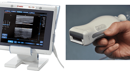
Diagnostic Ultrasound
Ultrasonography involves the use of reflected sound waves from tissue interfaces to draw an acoustic picture of a structure. Ultrasonic scanners are used in ophthalmology in two modes: A mode and B mode (also known as A scan and B scan, respectively). In A mode they measure the axial length of the eye. The eye measures between 21 mm and 26 mm in length. This measurement is used for calculating the power of the intraocular lens that should be implanted after the removal of a cataract. In B mode the ultrasound provides a 2-D image of the interior structures of the eye that permits detection of retinal detachments, foreign bodies, and tumors. This is especially useful when the light path of the eye is obstructed by a cloudy cataract or by blood in the vitreous, for instance, and viewing the interior of the eye cannot be accomplished using conventional optical instruments. Some of the most recent models of B scan machines have software that assembles 3-D images.
Ismael Cordero, CBET, is a health care technology specialist with ORBIS International, a nonprofit humanitarian organization dedicated to preventing blindness through training, institutional development, and public awareness.