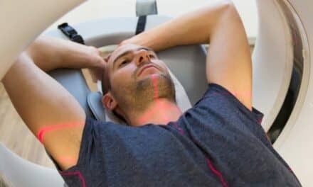
Summary: Hyperfine, in collaboration with the Medical University of South Carolina, will use the Swoop Portable MR Imaging system to scan astronauts’ brains before and after the Polaris Dawn mission to study changes in intracranial volume and understand the effects of spaceflight.
Key Takeaways:
- The Swoop system will provide the earliest brain images ever taken of astronauts immediately after returning to Earth.
- The research aims to uncover insights into spaceflight-associated neuro-ocular syndrome (SANS) and the brain’s adaptation to zero-gravity environments.
Hyperfine announced a collaboration with the Medical University of South Carolina to assess the brains of astronauts before and after embarking on the Polaris Dawn mission.
Use of Swoop Portable MR Imaging System
Researchers will utilize the Swoop Portable MR Imaging system at a SpaceX facility in Florida to acquire detailed images of astronauts’ brains within hours of their return from space following the Polaris Dawn mission. The research aims to use brain images acquired with the Swoop system to assess intracranial volume changes following spaceflight by performing a volumetric analysis of the brain and cerebrospinal fluid spaces. These scans will provide the earliest brain images ever taken of astronauts following their return to Earth.
“This research will help us understand how human physiology adapts to spaceflight and zero-gravity environments,” said Donna Roberts, MD, MS, principal investigator from the Medical University of South Carolina. “Using brain images acquired with the Swoop system, we aim to gain valuable insights into the etiology of spaceflight-associated neuro-ocular syndrome (SANS)—a condition in which astronauts experience changes in vision, alterations to the retina, and in some cases, swelling of the optic disc and increased intracranial pressure.”
Scan Timeline and Objectives
The Swoop system will acquire MR brain images of the four Polaris Dawn crewmembers at critical points before and after spaceflight—seven days before launch, within hours of return, and one day after return. These images will provide data to help researchers determine whether intracranial venous congestion occurs during spaceflight or during the brain’s re-adaptation to Earth.
“At Hyperfine, Inc., we are dedicated to enhancing brain health, and this research has the potential to advance neurological research,” said Maria Sainz, President and CEO of Hyperfine. “This collaboration showcases the versatility and robustness of our system and the potential of our groundbreaking technology to go into nearly any professional healthcare setting.”


