Patient empowerment, education, and engagement key focuses for today’s providers
By Elaine Sanchez Wilson
This past June, the American College of Obstetricians and Gynecologists (ACOG) released updated breast cancer screening recommendations, which were revised to emphasize “patient-provider shared decision-making” for average-risk women.
“Our new guidance considers each individual patient and her values,” says Christopher M. Zahn, MD, ACOG vice president of practice activities, in a statement. “Given the range of current recommendations, we have moved toward encouraging obstetrician–gynecologists to help their patients make personal screening choices from a range of reasonable options.”
According to Robert Fabrizio, director of strategic marketing for digital x-ray and women’s health at Stamford, Conn.-based Fujifilm Medical Systems U.S.A. Inc., the most successful practices already provide this level of care. In fact, a growing number of providers aim to engage their patients in personal discussions on breast density and risks, for example. Others may even explain imaging results before the patient leaves the office.
These efforts to bolster the patient-provider dialogue are meant to combat the confusion and fear associated with women’s experiences navigating a complex healthcare landscape.
“The challenges providers have in respect to mammography can oftentimes include dispelling misinformation about mammograms—including conflicting recommendations about when and how often to receive an exam—concerns about radiation and various invasive exam experiences associated,” Fabrizio says. “Many patients also fear discomfort from exams or may have firsthand experience with false positives, callbacks, or even biopsies.”
As a result, Fabrizio supports the ACOG’s focus on shared decisionmaking. “Mammography is very personal, so it’s no wonder that a patient will develop a loyalty and refer other patients to that imaging center if they have a consistently good experience throughout the entire screening and diagnostic process,” he adds.
Agreeing, Pam Cumming, senior director of product marketing for women’s health at Malvern, Pa.-based Siemens Healthineers, says she believes the ACOG guidelines represent a positive step toward fostering an important, close-knit relationship with a physician on many levels.
“Many women consider their Ob/Gyn as a gatekeeper for their health for many years—their reproductive years and then beyond for a lot of women,” she says. “Obviously, that relationship can be very personal and maybe even more so for some women than their general practitioners. It’s a very private and special relationship that is built and that can create a positive environment for discussion.”
Often, patients come to appointments with questions about what they’ve read online, Cumming observes. “In this world of the Internet and empowerment and the ability to take responsibility for your own health, the idea that this is a shared decision-making process is—for a very large segment of the demographic, I think—welcome and also comforting,” she says.
Individualized Care
Facilitating positive patient experiences is a top goal at Kansas City, Mo.-based Imaging for Women, a women’s imaging center that performs 16,700 exams annually. At Imaging for Woman, patients know their results at the end of their exams and receive their ‘lay letters’ when departing the office. The practice also reports exam results promptly to referring physicians, usually within an hour of the exam’s completion.
Mark J. Malley, MD, DABR, a physician at Imaging for Women, notes that a patient-centered care model does not necessarily align with efficiency; however, the tradeoff, he believes, is well worth it. “Although it is much more efficient to batch read, is it better for the patient to wait a few days or a week to get results or to have to come back for additional views? If I was a woman undergoing the exam, I [wouldn’t] think so!”
Patient-centric service can also translate into higher operational costs. “One must expend more resources to make a center patient-friendly and give same-day results,” Malley continues. “However, this is what distinguishes us from every hospital and every other imaging center…Our referral base knows that each woman will get a high-quality exam by someone who looks at the patient, examines the patient, and talks with the patient.”
The growing awareness that breast cancer screening needs to be tailored to the risk profile of the individual woman presents opportunities—and challenges, says Jonas Rehn, senior product manager of mammography at Philips Healthcare. Take, for instance, breast density level, which impacts the risk of developing breast cancer and raises questions on the sensitivity of mammography as a procedure to detect a growing lesion.
“In response to this awareness, we now see an increasing level of information being shared by healthcare providers with women about their breast density and their options to pursue adjunct screening methods,” Rehn continues. “There are multiple approaches for measuring breast density, which can cause challenges for the healthcare organizations, both in terms of additional time and effort spent subjectively assessing breast density. It also creates the risk of having the validity of the assessment questioned.”
According to Megan Rosengarten, vice president of global marketing at Marlborough, Mass.-based Hologic, significant resources are invested in driving educational initiatives, all in an effort to have women take an active role in their breast health.
The ultimate goal? To ensure that “patients and referring physicians alike are knowledgeable about the importance of breast cancer screening, the options available to them and how they best fit their need, and that they have an adequate understanding of the limitations of current risk assessment tools, which may not always include everything that influences a woman’s risk,” she says.
Choosing the Right Technology
One technology patients have been increasingly requesting is 3D mammography, reveals Dean J. Phillips, DO, president and director of outpatient imaging at New York-based Northern Radiology Imaging, PLLC. Fortunately for them, the law is on their side: Under New York Insurance Law, health insurers are now required to provide medically necessary coverage for 3D mammograms without co-pays, coinsurance, or deductibles.
With a commitment to offer the newest technology to women in the community—“a core value for us,” he adds—the practice searched for a system that would meet their patients’ needs and enable the business to stay competitive within the marketplace. “We decided to purchase Fujifilm’s 3D system based on the cost, our strong relationship with the local service technician, and ease of integration into our existing PACS technology,” Phillips says. Fujifilm’s Aspire Cristalle 3D digital mammography system was installed in July.
According to Fabrizio, Fujifilm’s system employs advanced automatic exposure control and image-based spectrum conversion, which uses a rapid feature recognition analysis of the breast structure to formulate optimal dose and image processing, automatically adjusting for individual areas of fatty and dense tissue, implants, chest wall, and skin line.
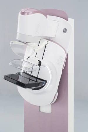
Meanwhile, at Missouri’s Imaging for Women, a search for a system that promotes patient comfort, quality imaging, and good service led the group to GE Healthcare’s Senographe Pristina mammography system. Two systems were installed in May and June, joining a GE Healthcare Senographe Essential with stereotactic capability installed in 2014.
“The key benefits of the Senographe Pristina system include better patient comfort, the fact that technologists find this system easy to use, its low dose, and its [production of] high-quality exams,” Malley says.
One of the most notable aspects of the Senographe Pristina is that the 3D mammography system delivers the same dose as 2D, according to Barbara Rhoden, USCAN region product marketing director for GE Healthcare Women’s Health. What’s more, during the Senographe Pristina’s development process, GE interviewed several patients to discuss their previous experiences with getting a mammogram, Rhoden reveals.
“Our design team learned that women complained about an uncomfortable examination due to pain experienced during compression,” she says. “Our team took these insights and expressly designed Senographe Pristina to revolutionize the mammography experience. The team designed a carbon Bucky—where you position the breast—which is warm to the touch, compared to traditional metallic approaches. This also helps reduce any pinching around the armpit during a mediolateral oblique examination.”
Women can now also lean their arms comfortably on dedicated armrests in order to help them relax during examinations, Rhoden adds. “We knew that women were nervous during the breast compression and that they were tensing their pectoral muscles while grabbing the hand grips,” Rhoden says. “This can potentially impact the quality of the images. So we changed that.”
Hologic’s Rosengarten says the company also solicits feedback from women, technologists, radiologists, and administrators regarding mammography. “In fact, Hologic was the first to bring breast tomosynthesis technology to the market in 2011 with the Genius 3D Mammography exam on the Selenia Dimensions system,” she says.
A key benefit of the Genius exam, according to Rosengarten, is its ability to detect 20% to 65% more invasive breast cancers compared to 2D alone. Furthermore, when compared to 2D mammography alone, the Genius 3D Mammography exam also reduces callbacks by up to 40%, she continues. And with Hologic’s Affirm prone breast biopsy system, she says, physicians are able to offer 3D imaging in the prone position—“the preferred positioning for many physicians as it offers a more compassionate biopsy experience without direct view of the needle.”
Philips, Jonas Rehn says, is also working to make personalized breast care a reality. Look no further than the company’s MicroDose SI low-dose mammography solution, he says, which can objectively measure breast density with a unique spectral imaging photon-counting technology. “The unit will produce a direct measure of density as part of the normal mammography procedure, without the need for additional exposures, hardware, or user intervention,” he says.
Still, Siemens’ Pam Cumming argues, Siemens is the only vendor that has FDA approval to diagnose from the 3D image set alone, eliminating the need for a 2D image. Specifically, the FDA has approved the use of 3D-only screening mammography utilizing the company’s Mammomat Inspiration with Tomosynthesis Option digital mammography system. To Cumming, this “a step forward” in early breast cancer detection.
What’s noteworthy about 3D breast tomosynthesis, Cumming says, is that it creates slices of the breast, which enable radiologists to scroll through the breast volume.
“The data that is created during the [tomosynthesis] scan is reprocessed using sophisticated algorithms, designed to reduce noise and enhance tissue morphology—subtle distortions in architecture, where small cancers are more evident,” she says. “Oftentimes, there can be micro-calcifications in veins that, under this 3D image volume rendering from Siemens, you can actually see where they are in a 3D space.” Such technological advancements can help clinicians facilitate better patient care, she maintains.
The Future of Women’s Health
Cumming says she anticipates a growing adoption of 3D breast tomosynthesis, along with more consumer awareness around the technology. “More women are asking for it, and that’s helping to drive the conversion.” She also believes there will continue to be more discussion surrounding the recommended frequency of screening for different subsets of women. While some guidelines recommend mammograms every two years, she points to research suggesting that cancers may happen in the off-year.
“When that happens and you can get something early, you might save a woman the ordeal of chemo or radiation,” Cumming says. “I think we’re going to continue to see strong voices on annual screening from the governing bodies, for the most part.”
Rosengarten agrees. “We know that women will continue to demand the most accurate exam, and 3D mammography will continue to establish itself as the industry ‘gold standard’ for breast cancer screening…,” Rosengarten says. “We also predict we’ll continue to witness the rise of consumerism in the industry as patients realize more than ever the very real power they have to make informed, educated choices about their healthcare options.”
With an eye toward the future, GE’s Barbara Rhoden perhaps sums it up best, however, saying: “We believe that we are entering an era where a combination of patient experience, superior diagnostic accuracy, and low dose is driving the adoption of mammography equipment.” And to provide the best possible patient care, Rhoden says, one must deliver all three.
Elaine Sanchez Wilson is associate editor of 24×7 Magazine. For more information, contact [email protected].
SIDEBAR: Summary of ACOG’s Updated Recommendations for Screening Mammography
• Women at average risk of breast cancer should be offered screening mammography starting at age 40 years. If they have not initiated screening in their 40s, they should begin screening mammography by no later than age 50. The decision about the age to begin mammography screening should be made through a shared decision-making process. This discussion should include information about the potential benefits and harms.
• Women at average risk of breast cancer should have screening mammography every one or two years based on an informed, shared decision-making process that includes a discussion of the benefits and harms of annual and biennial screening and incorporates patient values and preferences.
• Women at average risk of breast cancer should continue screening mammography until at least 75 years. Beyond age 75, the decision to discontinue screening mammography should be based on a shared decision-making process informed by the woman’s health status and longevity.
—The American College of Obstetricians and Gynecologists


