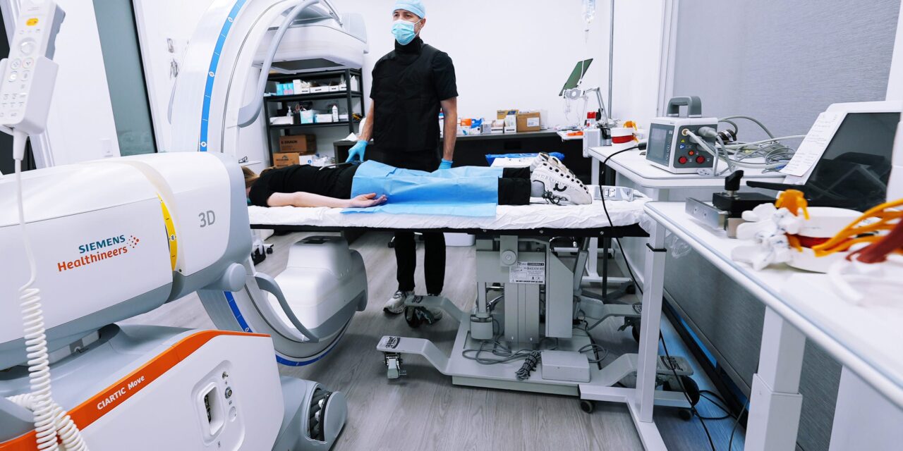
The imaging system includes self-driving robotic guidance with automated positioning to support minimally invasive procedures.
Alpine Spine & Orthopedics Institute has implemented the new Ciartic Move 3D imaging system, an advanced imaging system that provides ultra-high-resolution 3D imaging with robotically guided movements, overcoming many of the limits of 2D image slices and using self-navigating capabilities to reduce the need for manual operation while also improving workflow during orthopedic procedures.
The Siemens 3D Ciartic Move is a low-profile mobile imaging device that enables 3D navigation with robotic holonomic mechanisms to allow floating-like movements around a surgical field.
“This technology really moves us into the future for minimally-invasive image-guided procedures,” says Richard J. McMurtrey, MD, MSc, in a release. “It is the most advanced robotic 3D C-arm cone beam scanner that exists for interventional procedures to see targets and approaches in the highest resolution, and we are grateful to be the first in Utah to implement a revolutionary device of this kind.”
The Ciartic mobile 3D imaging system introduces a mobile imaging solution and advances intricate and complex minimally invasive procedures and interventions in spine, orthopedics, and trauma by bringing together advanced technologies from 3D computed tomography with 3D navigation and needle guidance, metal artifact reduction and hardware detection, high-resolution fluoroscopy and magnification, digital subtraction angiography, low-dose radiation settings, and self-driving robotic guidance with automated positioning all at the touch of a button.
“As both a doctor and an engineer, it is rewarding to lead the way in combining the best of these fields to provide the most advanced and most minimally invasive care possible,” says McMurtrey in a release.
The clinic has already implemented the system in numerous procedures, gaining new insights into the nuances of each injury and unique perspectives in minimally invasive approaches to the critical features and pain triggering points of each injury, including spinal pars defects, sacroiliac dysfunction, spino-sacral anomalies, cranio-cervical instabilities, ligament destabilization, cartilage injuries and arthritic joint pathologies, degenerative disc defects and facet arthropathy affecting nerve roots and branches.
Improved Imaging Reveals Complex Injuries
In the first week of implementing the system, it enabled clear visualization of several complex problems that MRI imaging had been unable to fully clarify, according to a release from the company, including stress fractures in the spine and non-union fractures of the scaphoid bone in the wrist and of the sesamoid, metatarsal, and navicular bones in the foot, as well as complex bone spurs, synovial cysts, and subchondral bone cysts with arthritis that can be patched with orthobiologic scaffolding including patient’s own platelet-rich fibrin, bone marrow stem cells, and other regenerative agents.
Furthermore, this technology helps confirm needle placement in small joints such as the atlanto-axial joints, atlanto-occipital joints, and facet joints of the spine.
“We focus on repairing the underlying injuries as directly as possible with minimally invasive image-guided procedures that let us see and target many types of orthopedic and neurologic problems from every angle,” says McMurtrey in a release.
McMurtrey explains that many patients have painful conditions that surgery cannot address at all, or issues where surgery can do more harm than good, in addition to the many patients who have already undergone spine fusions or joint surgeries yet still continue to have significant pain and dysfunction.
“The Ciartic Move device is a powerful tool to help us find and target those sources of pain and directly treat a variety of injuries with minimally invasive techniques that promote long-term recovery and more regenerative options to help repair tissue injuries and defects directly, and in this way, this device and approach are really leading us into the future,” he says in a release.
Summary:
Alpine Spine & Orthopedics has implemented the Ciartic Move 3D imaging system, a robotic, high-resolution imaging device for use in spine and orthopedic procedures. The system provides 3D navigation, automated positioning, and advanced imaging capabilities, offering a more detailed view of complex injuries and surgical targets. According to Alpine Spine & Orthopedics, the imaging system has already been used in multiple procedures, providing new perspectives on spinal defects, joint pathologies, and fractures that were not fully visible on MRI scans. The clinic is the first in Utah to integrate the technolog.
Key Takeaways:
- Alpine Spine & Orthopedics Introduces New 3D Imaging System – The clinic has adopted the Ciartic Move 3D system, which provides robotic navigation and high-resolution imaging.
- New Imaging Capabilities Offer Additional Diagnostic Insights – The system has helped identify fractures, cysts, and bone defects that were not fully visible on MRI scans, according to the clinic.
- First Implementation of the System in Utah – Alpine Spine & Orthopedics is the first in the state to integrate the Ciartic Move 3D imaging system into its surgical workflow.
Photo caption: Ciartic Move 3D imaging system at Alpine Spine & Orthopedics Institute
Photo credit: Alpine Spine & Orthopedics Institute


