 Siemens Healthcare has received clearance from the FDA for its Artis Q and Artis Q.zen angiography system families, which feature new x-ray tube and detector technology designed to improve minimally invasive therapy of diseases such as coronary artery disease (CAD), stroke, and cancer.
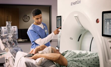
The new x-ray tube in both the Artis Q (pictured) and Artis Q.zen can help physicians identify small vessels. The Artis Q.zen combines this x-ray source with a new detector technology that supports interventional imaging in ultra-low-dose ranges to patients, physicians, and medical staff—particularly during longer interventions.
In lieu of the coiled filaments found in conventional x-ray tubes, Siemens uses second-generation flat emitter technology in the new x-ray tube of the Artis Q and Artis Q.zen product lines. These flat emitters enable smaller quadratic focal spots that lead to improved visibility of small vessels by as much as 70% and a high level of detailed imaging information, according to Siemens.
The Artis Q.zen system family combines the new x-ray tube with a new detector technology that enables detection at ultra-low radiation levels. Artis Q.zen imaging can utilize doses as low as half the standard levels applied in angiography. The company stated that this improvement is the result of several innovations, including a fundamental change in detector design from amorphous silicon to a more homogenous crystalline silicon structure. This structure allows for more effective signal amplification, greatly reducing electronic noise even at ultra-low radiation doses.
In addition to these hardware innovations, the Artis Q and Artis Q.zen system families possess software applications designed to improve interventional imaging. In the treatment of CAD, the new integrated intravascular ultrasound (IVUS) map application automatically and precisely co-registers IVUS images and angiography images, adding detailed IVUS data such as vessel, lumen, and wall structure. Additionally, CLEARstent Live allows the cardiologist to image stents with motion stabilization in real time by freezing motion in the region defined by the balloon markers.