Improved radioactive isotopes and new hybrid systems allow for the earlier detection of disease and more targeted treatment
 When it comes to medical imaging modalities, computed tomography (CT), radiology, angiography, cardiac catheterization, magnetic resonance imaging (MRI), and ultrasound spring most readily to mind. Often overlooked is nuclear medicine.
Not as visible as most other imaging modalities, nuclear medicine involves the use of radioactive isotopes and was actually in existence as an imaging modality prior to the arrival of ultrasound or CT. While most other imaging modalities are primarily used for diagnosis, nuclear medicine is used for both the diagnosis and treatment of disease.
In nuclear medicine’s formative years, radioactive materials were used mostly to treat various forms of cancer. It was not widely used as an imaging modality until the 1950s, when scanners and special cameras were developed to detect the presence of radioactive materials in the body. Archaic in comparison to today’s equipment, these early nuclear medicine systems resulted in lengthier and more tedious procedures that used higher doses of radioisotopes.
Don Gouger, CNMT, radiology director at Lee Memorial Hospital in Fort Myers, Fla, has been involved with nuclear medicine since its infancy. With 40 years of experience, Gouger recalls that the first nuclear medicine imaging device he worked with was known as a rectilinear scanner. “This scanner scanned one line at a time, kind of like a typewriter,” Gouger says. “In those days, a whole-body bone scan would take almost all day.” Today, those same scans take approximately 1 hour.
Some of the procedures performed by CT or ultrasound today, such as brain scans, were originally performed with nuclear medicine equipment. “In those days, we were doing 10 to 12 brain scans per day, which took 1 to 2 hours per scan,” Gouger says.
With more than 100 different nuclear medicine procedures now available, nuclear medicine is used to treat conditions such as thyroid cancer and hyperthyroidism; and for detecting tumors, aneurysms, inadequate blood flow, and blood-cell disorders. The major improvements in equipment and in radioactive isotopes have made nuclear-medicine imaging both safer and faster.
Nuclear medicine differs from other imaging modalities in a number of ways. Unique in its ability to diagnose bodily function at the molecular level, nuclear medicine looks at the function of body parts while most other modalities look at the anatomical position of those parts.
For example, a CT procedure might show the existence of brain tissue (dead or alive), while a nuclear medicine procedure would show only the brain tissue that is alive (or has blood flow), according to John Stuivenga, CBET, clinical engineering account manager at Catholic Health Initiative (CHI) hospitals in the Seattle area.
Another major difference between nuclear medicine and other imaging modalities is its source of radiation. All imaging modalities that rely on radiation for imaging generate their own radiation, except for nuclear medicine. In nuclear medicine, the patient gives off the radiation by way of injected or ingested radioactive isotopes. These isotopes have varying levels of radioactive properties; and different types of isotopes and isotope solutions are used, depending upon the part of the anatomy to be imaged. Different isotopes tend to concentrate in particular organs. For example, iodine-131 settles in the thyroid gland and can reveal defects in thyroid functioning, while the isotope carbon-14 can be helpful in studying metabolism abnormalities that may reveal diabetes.
The Hazards
Hazards in the nuclear medicine environment range from radioactive hazards to mechanical hazards. In most modalities, radiation safety concerns center on the radiation produced by the machine during the procedure. In nuclear medicine, the concerns relate to the storage and handling of radioactive isotopes, which is tightly regulated, according to Tim Zinsmeister, RTN, CNMT, nuclear medicine manager at Akron, Ohio’s Summa Health System. “Radiopharmaceuticals, used in nuclear medicine, typically have a half-life anywhere between 2 hours and 8 days,” Zinsmeister says. While these substances are low dose and have a relatively short half-life, it is important to be aware of their existence and dangers. Protective clothing and monitoring devices are used to help prevent or detect exposure.
One major concern in the handling of these substances is the possibility of contamination of the equipment. Contamination can cause false readings during calibrations or patient studies, and it presents a danger of unwanted exposure. Nuclear medicine technicians and biomeds use a radiation survey meter to check the area and clothing for cross contamination and take appropriate action if any is found.
The Systems
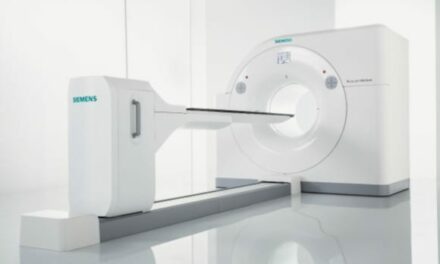
Positron emission tomography (PET), single photon emission computed tomography (SPECT), cardiovascular imaging, bone scanning, and thyroid uptake are the main procedures and/or equipment used in nuclear medicine studies. The basic nuclear medicine systems include a single- or dual-camera head (or detector), a gantry (or housing), a patient table, lead collimators, monitors (called p-scopes), and a processing station. These systems perform different types of nuclear medicine studies or procedures.
Dedicated PET scanners have a doughnut-shaped gantry—much like a CT or MRI gantry. The gantry contains the circular gamma ray detector array, and the patient is moved through the gantry via a patient table.
The hybrid PET/CT system has been around for several years, but it has recently become more predominant, according to Zinsmeister. It combines PET and CT technology in a single gantry, creating a more accurate diagnosis. This combination helps to positively identify problems without the need to move the patient. “The patient can get the CT and, while lying in the same position, receive the PET scan. Then the CT and PET images are merged to aid in diagnosis, especially since the patient is in the same exact orientation and position,” Zinsmeister says.
Another hybrid—the SPECT/CT system—also allows the patient to remain in the same position for both scans. The SPECT/CT system greatly enhances anatomical mapping and localization, and the latest SPECT tracers are more targeted, seeking only the tissues they’ve been designed to find.
The heavy mechanical moving parts of a nuclear medicine system, such as the tables, gantries, and collimators, can present hazards to those who use or service the equipment.
“The gantries have large gears and powerful motors. You don’t want to get hit or caught by these moving parts,” says Gene Hollowell, CE, field service specialist at CHI Penrose Hospital, Colorado Springs, Colo.
The detector is composed of several components, including a sodium crystal surface, photomultiplier tubes (PMT), and the electronics used to convert the PMT signals to image data. The radiation emitted by a patient passes through a lead collimator, strikes the sodium crystal surface—which produces light that is seen by the PMTs—and is then converted to digital data. This data is manipulated in the processing stations to provide diagnostic images for physicians.
The collimators are heavy lead devices that go between the patient and the detector, and are commonly attached to the detector assembly via mounting surfaces and mechanical latches. These collimators typically have an internal structure that is in the shape of a honeycomb or a venetian blind, depending upon the application.
The detector must be checked for uniformity on a daily basis. “Uniformity is the process of making the detector look homogeneous,” says John Roberts, a biomed imaging/IT specialist at Fort Myers-based Lee Memorial Health System. Uniformity checks require the use of a radioactive source. This source radiates the detector, and tests are done to verify the uniformity. If uniformity cannot be obtained, then a calibration is most likely needed.
During semiannual preventive maintenance (PM), nuclear medicine service engineers check detectors for proper calibration. Calibrating a detector array can be very time consuming, taking 4 to 8 hours, according to Roberts. “Some calibrations require you to take the back cover off of the head, which can create temperature drift and heat issues, and thus lengthen the calibration time.”
Roberts points out that there are basically five different calibrations that need to be performed to tune the detector: PMT adjustment, offset, linearity, energy, and uniformity. “The uniformity calibration itself is specific to each isotope used,” Roberts says. “If you use five different isotopes, you may need to do five different uniformity calibrations.”
“Mechanical devices such as latches, motors, gantries, tables, and brakes all need to be adjusted, cleaned, or lubricated during PMs,” Stuivenga says.
Checking the mechanical devices and performing electrical checks can take the better part of a day, according to Hollowell, who tries to break these PMs into one major mechanical PM and one major electrical PM per year.
What Could Go Wrong?
Common failure items in a nuclear medicine system include latches, p-scopes, mechanical parts, and power supplies. Other items that are not as likely to fail are PMT tubes, electronic circuitry, and crystals, according to Stuivenga. “A crystal can cost tens of thousands of dollars,” he says.
PMT tube failures occur more often than crystals, according to Roberts, who has changed only one crystal in his 14-plus years of experience. “The time to replace a bad PMT varies,” Roberts says. “Most cameras could have 40 to 90 PMTs in each head. If one goes bad, you have to take all of the electronics out in order to replace it.”
Some of the challenges a nuclear medicine service engineer faces, such as patient care needs, are similar to those in other imaging modalities. A room might need repair, but it may still be functioning well enough for the procedures to continue. In circumstances when patients are scheduled, repairs must wait until the room is available.
Another challenge is the inability to perform calibrations while patients are being scanned on other nuclear medicine equipment in the area. “If the walls between equipment are not lead lined and patients are being scanned in the area, your calibration will be affected,” Stuivenga says. “The patient is much ‘hotter’ than the small source used for calibration, so the detector you are calibrating will pick up their radiation and give you a false reading.” Both of these situations often create overtime, after-hours, and weekend work.
On the Horizon
As hospitals constantly face the dilemma of rising costs coupled with decreased revenue, many have lowered costs by reducing their number of service contracts, creating opportunities for biomeds. “We rely on biomeds quite a bit,” Zinsmeister says. “They help to reduce the downtime that would occur when waiting on the vendor.”
“Eliminating or modifying those contracts to a ‘first look’ type of contract can greatly reduce the cost and can provide justification to hire or promote a biomed into that area of responsibility,” Hollowell says.
Stuivenga knows how in-house service and training can pave the way for new opportunities. He got his own start in nuclear medicine service in 1990 after his manager asked him to replace a board in one of their nuclear medicine systems. Impressed with his work, the nuclear medicine supervisor requested him for future service. Soon after, Stuivenga’s hospital purchased new nuclear medicine equipment along with technical training and he was asked to attend the course. His suggestion: “Keep your eyes open, be eager and willing to get involved, and present yourself well.”
Mike Adams is a contributing writer for 24×7.