The catheter is designed to offer a clearer view of the heart during procedures to treat abnormal heart rhythms.
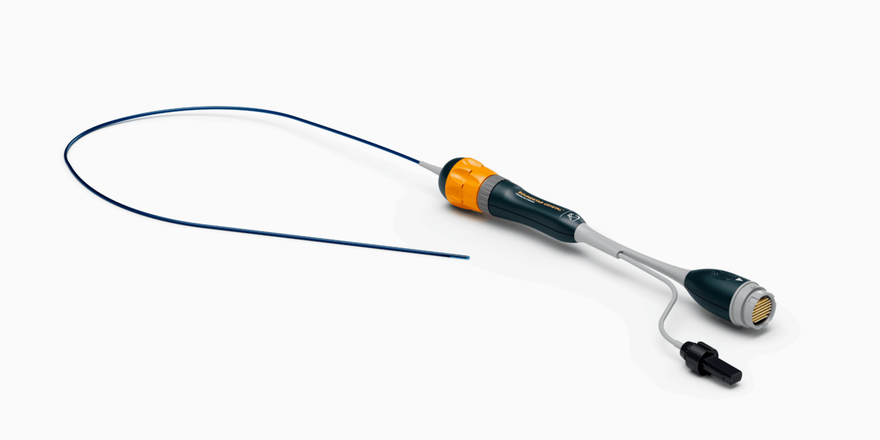
Johnson & Johnson MedTech has announced the US launch of its Soundstar Crystal ultrasound catheter, designed to provide enhanced intracardiac echocardiography (ICE) imaging during cardiac ablation procedures.
The 2D ICE catheter offers improved image quality compared to previous ICE devices and integrates with the company’s Carto 3 Mapping System, including the CartoSound Fam module. Powered by an artificial intelligence algorithm, CartoSound Fam enables automated generation of left atrial anatomy using images captured from ultrasound catheter rotation in the right atrium. This integration is intended to streamline the mapping workflow for electrophysiologists treating cardiac arrhythmias.
“During the cases I performed with the Soundstar Crystal ultrasound catheter, I was impressed by the clear visualization, tissue definition, enhanced far-field imaging, and full integration with other platforms,” says Amin Al-Ahmad, MD, St. David’s HealthCare, Austin, TX.
ICE is widely used during catheter ablation procedures to visualize cardiac structures in real time, monitor catheter-tissue contact, and identify potential complications. When paired with 3D electroanatomical mapping systems like Carto 3, ICE imaging can support zero-fluoroscopy procedures and allow for ablations under conscious sedation without the need for esophageal intubation.
The Soundstar Crystal catheter joins Johnson & Johnson MedTech’s broader ultrasound portfolio, which also includes the NuVision catheter for 4D cardiac imaging.
According to the company, the growing prevalence of atrial fibrillation—which affects more than 50 million people globally and over 8 million in the US—continues to drive demand for ablation tools. Left untreated, AFib can progress and lead to complications such as heart disease or stroke.
Photo caption: Soundstar Crystal ultrasound catheter
Photo credit: Johnson & Johnson MedTech